投稿系统
投稿系统


LncRNA MIR4435-2HG promotes psoriatic inflammation via activation of PDGFRB-positive skin mesenchymal cells
doi: 10.1515/fzm-2026-0012
-
Abstract:
Background Psoriasis is a chronic inflammatory skin disease influenced by genetic, immune, and environmental factors. Notably, its prevalence is significantly higher in colder regions, and cold climates are known to exacerbate disease symptoms. This study aimed to investigate the role of the long non-coding RNA MIR4435-2HG in regulating inflammation and cytokine production in psoriatic skin. Methods Single-cell RNA sequencing identified MIR4435-2HG expression in PDGFRB-positive mesenchymal cell subsets in psoriatic skin. Functional assays were performed to evaluate its effects on inflammatory cytokine expression in mesenchymal cells activated by TNF-α. An imiquimod-induced psoriasis mouse model was used to assess the effects of MIR4435-2HG knockdown in vivo. Results MIR4435-2HG was found to upregulate IL-6, CXCL8, and IL-17B expression in TNF-α-activated mesenchymal cells. Knockdown of MIR4435-2HG in the mouse model significantly reduced skin inflammation, epidermal hyperplasia, and psoriasis-associated cytokine expression. Conclusion MIR4435-2HG plays a critical role in regulating inflammation through mesenchymal cell activation, offering a potential therapeutic target for reducing psoriasis severity, especially in cold climates. -
Key words:
- psoriasis /
- mesenchymal cell /
- MIR4435-2HG
-

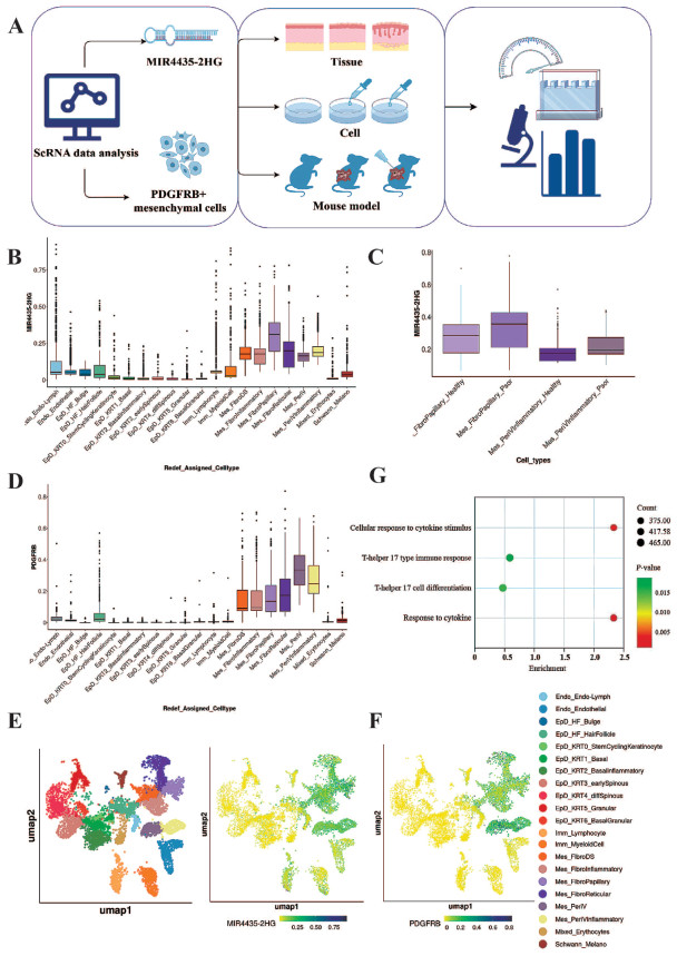
Figure 1. Single-cell RNA sequencing data reveal specific expression of MIR4435-2HG in PDGFRB-positive mesenchymal cell clusters
(A) Schematic workflow illustrating the experimental design and analytical pipeline used in this study, designed by Figdraw2.0 (Home for Researchers, Hangzhou, China). (B) Box plot showing the relative expression of MIR4435-2HG across different skin cell clusters following updated classification. The x-axis represents cell cluster identities, and the y-axis indicates the relative expression level of MIR4435-2HG. Clusters selected for further investigation are highlighted in red. (C) Box plot comparing MIR4435-2HG expression between healthy and psoriatic mesenchymal cell clusters. (D) Box plot showing the relative expression of PDGFRB across different skin cell clusters. The x-axis represents cell cluster identities, and the y-axis indicates the relative expression level of PDGFRB. (E, F) UMAP plots illustrating the co-expression patterns of MIR4435-2HG and PDGFRB in skin cells. A substantial overlap in expression is observed within mesenchymal cell clusters. (G) Gene Ontology (GO) enrichment analysis of MIR4435-2HG-associated genes, highlighting their involvement in cellular responses to cytokine stimuli and cytokine signaling pathways.

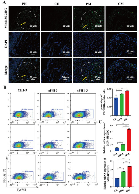
Figure 2. Comparison of MIR4435-2HG expression in PDGFRB-positive mesenchymal cells between psoriatic and healthy skin
(A) Fluorescence in situ hybridization (FISH) analysis showing MIR4435-2HG expression (green) in human and mouse healthy and psoriatic skin. DAPI (blue) indicates nuclear staining. Yellow arrowheads indicate MIR4435-2HG-positive foci, and white dashed circles denote high-expression regions. Scale bar: 50 μm. N = 3 biological replicates. (B) Flow cytometry analysis of PDGFRB-positive mesenchymal cell proportions in skin tissues from patients with different psoriasis severities. Tyr751 represents PDGFRB-positive signals, FSC-A represents cell size, and the blue box indicates the proportion of PDGFRB-positive cells. N = 3 biological replicates. (C) Quantitative PCR (qPCR) analysis showing relative MIR4435-2HG mRNA expression in PDGFRB-positive cells isolated from healthy (CM/CH), mild psoriasis (mPM/mPH), and moderate-to-severe psoriasis (sPM/sPH) skin in both mice and humans. **P < 0.01; N = 9 biological replicates. CH, human healthy control; CM, mouse healthy control; PH, human psoriasis; PM, mouse psoriasis; mPH, mild psoriasis; sPH, moderate-to-severe psoriasis.

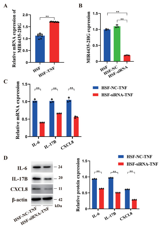
Figure 3. MIR4435-2HG regulates psoriasis-related cytokine expression in TNF-α-induced dermal mesenchymal cells
(A) Quantitative PCR (qPCR) analysis showing relative MIR4435-2HG mRNA expression in healthy dermal fibroblasts (HSF) and TNF-α-treated HSF cells (HSF-TNF). Data are presented as mean ± SEM. P < 0.01; N = 3 biological replicates. (B) qPCR validation of MIR4435-2HG knockdown in HSF cells using RNA interference (HSF-siRNA-TNF). The HSF-NC-TNF group served as a negative control. Data are presented as mean ± SEM. P < 0.01; N = 3 biological replicates. (C, D) qPCR and Western blot analyses showing the effects of MIR4435-2HG silencing on TNF-α-induced mRNA and protein expression of interleukin (IL)-6, IL-17B, and CXCL8 in HSF cells. Band intensities were normalized to β-actin. Molecular weight markers are indicated in kDa (kilodaltons). Data are presented as mean ± SEM. **P < 0.01; N = 3 biological replicates.

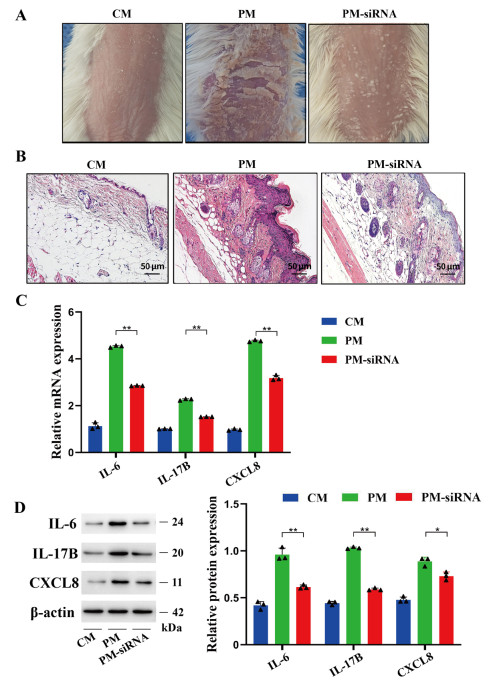
Figure 4. Suppressing MIR4435-2HG expression alleviates psoriasis severity in a murine model
(A, B) Representative images of dorsal skin and hematoxylin and eosin (H&E)-stained sections from healthy mice (CM), psoriasis model mice (PM), and siRNA-treated psoriasis mice (PM-siRNA). (C, D) Quantitative PCR (qPCR) and Western blot analyses showing the expression levels of interleukin (IL)-6, IL-17B, and CXCL8 in dermal cells isolated from dorsal skin across the three groups. Bar = 50 μm. Band intensities were normalized to β-actin. Molecular weight markers are indicated in kDa (kilodaltons). **P < 0.01; N = 3 biological replicates.

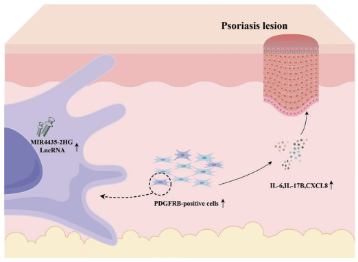
Figure 5. Mechanistic diagram illustrating the role of MIR4435-2HG in psoriasis pathogenesis
A schematic representation summarizing how MIR4435-2HG contributes to psoriasis pathogenesis through the activation of mesenchymal cells and the regulation of pro-inflammatory cytokine expression, including interleukin (IL)-6, IL-17B, and CXCL8. Designed by Figdraw2.0 (Home for Researchers, Hangzhou, China).
-
[1] Parisi R, Iskandar I Y K, Kontopantelis E, et al. National, regional, and worldwide epidemiology of psoriasis: Systematic analysis and modelling study. BMJ, 2020; 369: m1590. [2] Jensen K K, Serup J, Alsing K K. Psoriasis and seasonal variation: A systematic review on reports from Northern and Central Europe-Little overall variation but distinctive subsets with improvement in summer or wintertime. Skin Res Technol, 2022; 28(1): 180-186. doi: 10.1111/srt.13102 [3] Woolf R, Nussbaumer O, Hayday A. Potential for innate-like responsiveness of resident T cells in human skin: a new perspective on tissue immune-surveillance. The Lancet, 2016; 387: S108. doi: 10.1016/S0140-6736(16)00495-5 [4] Li Z, Cao T, Li Q, et al. Cross-disease characterization of fibroblast heterogeneities and their pathogenic roles in skin inflammation. Clin Immunol, 2023; 255: 109742. doi: 10.1016/j.clim.2023.109742 [5] Castillo R L, Sidhu I, Dolgalev I, et al. Spatial transcriptomics stratifies psoriatic disease severity by emergent cellular ecosystems. Sci Immunol, 2023; 8(84): eabq7991. doi: 10.1126/sciimmunol.abq7991 [6] Guo D, Li X, Wang J, et al. Single-cell RNA-seq reveals keratinocyte and fibroblast heterogeneity and their crosstalk via epithelialmesenchymal transition in psoriasis. Cell Death Dis, 2024; 15(3): 207. doi: 10.1038/s41419-024-06583-z [7] Gupta R, Ahn R, Lai K, et al. Landscape of long noncoding RNAs in psoriatic and healthy skin. J Invest Dermatol, 2016; 136(3): 603-609. doi: 10.1016/j.jid.2015.12.009 [8] Lin J, Li X, Zhang F, et al. Transcriptome wide analysis of long non-coding RNA-associated ceRNA regulatory circuits in psoriasis. J Cell Mol Med, 2021; 25(14): 6925-6935. doi: 10.1111/jcmm.16703 [9] Jiang Z, Feng T, Lu Z, et al. PDGFRb(+) mesenchymal cells, but not NG2(+) mural cells, contribute to cardiac fat. Cell Rep, 2021; 34(5): 108697. doi: 10.1016/j.celrep.2021.108697 [10] Zhang M, Yu X, Zhang Q, et al. MIR4435-2HG: A newly proposed lncRNA in human cancer. Biomed Pharmacother, 2022; 150: 112971. doi: 10.1016/j.biopha.2022.112971 [11] Cai S, Fan C, Xie L, et al. Single-cell RNA sequencing reveals the potential mechanism of heterogeneity of immunomodulatory properties of foreskin and umbilical cord mesenchymal stromal cells. Cell Biosci, 2022; 12(1): 115. doi: 10.1186/s13578-022-00848-w [12] Committee on Psoriasis, Chinese Society of Dermatology. Guideline for the diagnosis and treatment of psoriasis in China (2023 edition). Chinese Journal of Dermatology, 2023; 56(7): 573-625. [13] He Y, Huang M, Wang Y, et al. Inhibition of CTSC contributes to psoriasis inflammation and keratinocyte hyperproliferation by NF-κB signaling pathway. Int Immunopharmacol, 2025; 157: 114808. doi: 10.1016/j.intimp.2025.114808 [14] Kulig P, Brazauskas P, Suffiotti M, et al. Efficacy of IDOR-1117-2520, a novel, orally available CCR6 antagonist in preclinical models of skin dermatitis. Br J Pharmacol, 2025; 182(15): 3452-3475. doi: 10.1111/bph.70025 [15] Gao Y, Yao X, Zhai Y, et al. Single cell transcriptional zonation of human psoriasis skin identifies an alternative immunoregulatory axis conducted by skin resident cells. Cell Death Dis, 2021; 12(5): 450. doi: 10.1038/s41419-021-03724-6 [16] Kirkwood P M, Gibson D A, Shaw I, et al. Single-cell RNA sequencing and lineage tracing confirm mesenchyme to epithelial transformation (MET) contributes to repair of the endometrium at menstruation. Elife, 2022; 11: e77663. doi: 10.7554/eLife.77663 [17] Yoshida N, Yamamoto S, Hamashima T, et al. Dysregulation of Amphiregulin stimulates the pathogenesis of cystic lymphangioma. Proc Natl Acad Sci U S A, 2021; 118(19): e2019580118. doi: 10.1073/pnas.2019580118 [18] Li Y, He Y, Yang F, et al. Gasdermin E-mediated keratinocyte pyroptosis participates in the pathogenesis of psoriasis by promoting skin inflammation. Br J Dermatol, 2024; 191(3): 385-396. doi: 10.1093/bjd/ljae179 [19] Zhang S, Zhang J, Yu J, et al. Hyperforin ameliorates imiquimod-induced psoriasis-like murine skin inflammation by modulating IL-17A-producing γδ T cells. Front Immunol, 2021; 12: 635076. doi: 10.3389/fimmu.2021.635076 [20] Ma C, Gu C, Lian P, et al. Sulforaphane alleviates psoriasis by enhancing antioxidant defense through KEAP1-NRF2 Pathway activation and attenuating inflammatory signaling. Cell Death Dis, 2023; 14(11): 768. doi: 10.1038/s41419-023-06234-9 [21] Guo D, Wang Q, Li A, et al. Liquiritin targeting Th17 cells differentiation and abnormal proliferation of keratinocytes alleviates psoriasis via NF-κB and AP-1 pathway. Phytother Res, 2024; 38(1): 174-186. doi: 10.1002/ptr.8038 [22] Arican O, Aral M, Sasmaz S, et al. Serum levels of TNF-alpha, IFN-gamma, IL-6, IL-8, IL-12, IL-17, and IL-18 in patients with active psoriasis and correlation with disease severity. Mediators Inflamm, 2005; 2005(5): 273-279. doi: 10.1155/MI.2005.273 [23] Xu H, Liu J, Niu M, et al. Soluble IL-6R-mediated IL-6 trans-signaling activation contributes to the pathological development of psoriasis. J Mol Med (Berl), 2021; 99(7): 1009-1020. doi: 10.1007/s00109-021-02073-3 [24] Wu P, Ma G, Zhu X, et al. Cyr61/CCN1 is involved in the pathogenesis of psoriasis vulgaris via promoting IL-8 production by keratinocytes in a JNK/NF-κB pathway. Clin Immunol, 2017; 174: 53-62. doi: 10.1016/j.clim.2016.11.003 [25] Goodman W A, Levine A D, Massari J V, et al. IL-6 signaling in psoriasis prevents immune suppression by regulatory T cells. J Immunol, 2009; 183(5): 3170-3176. doi: 10.4049/jimmunol.0803721 [26] Lauffer F, Eyerich K, Boehncke W H, et al. Cytokines of the IL-17 family in psoriasis. J Dtsch Dermatol Ges, 2020; 18(7): 675-681. [27] Qazi B S, Tang K, Qazi A. Recent advances in underlying pathologies provide insight into interleukin-8 expression-mediated inflammation and angiogenesis. Int J Inflam, 2011; 2011: 908468. [28] van der Fits L, Mourits S, Voerman J S, et al. Imiquimod-induced psoriasis-like skin inflammation in mice is mediated via the IL-23/IL-17 axis. J Immunol, 2009; 182(9): 5836-5845. doi: 10.4049/jimmunol.0802999 [29] Gangwar R S, Gudjonsson J E, Ward N L. Mouse models of psoriasis: A comprehensive review. J Invest Dermatol, 2022; 142(3 Pt B): 884-897. [30] Muntyanu A, Milan R, Kaouache M, et al. Tree-based machine learning to identify predictors of psoriasis incidence at the neighborhood level: A populational study from Quebec, Canada. Am J Clin Dermatol, 2024; 25(3): 497-508. doi: 10.1007/s40257-024-00854-3 [31] Isler M F, Coates S J, Boos M D. Climate change, the cutaneous microbiome and skin disease: Implications for a warming world. Int J Dermatol, 2023; 62(3): 337-345. doi: 10.1111/ijd.16297 [32] Ma F, Plazyo O, Billi A C, et al. Single cell and spatial sequencing define processes by which keratinocytes and fibroblasts amplify inflammatory responses in psoriasis. Nat Commun, 2023; 14(1): 3455. doi: 10.1038/s41467-023-39020-4 [33] Kim J, Lee J, Kim H J, et al. Single-cell transcriptomics applied to emigrating cells from psoriasis elucidate pathogenic versus regulatory immune cell subsets. J Allergy Clin Immunol, 2021; 148(5): 1281-1292. doi: 10.1016/j.jaci.2021.04.021 [34] Xu Z, Chen D, Hu Y, et al. Anatomically distinct fibroblast subsets determine skin autoimmune patterns. Nature, 2022; 601(7891): 118-124. doi: 10.1038/s41586-021-04221-8 [35] Chen Z, Guan D, Zhu Q, et al. Biological roles and pathogenic mechanisms of LncRNA MIR4435-2HG in cancer: A comprehensive review. Curr Issues Mol Biol, 2023; 45(11): 8864-8881. doi: 10.3390/cimb45110556 [36] Wang X, Ren L, Chen S, e al. Long non-coding RNA MIR4435-2HG/microRNA-125a-5p axis is involved in myocardial ischemic injuries. Bioengineered, 2022; 13(4): 10707-10720. doi: 10.1080/21655979.2022.2051259 [37] Xiao Y, Bao Y, Tang L, et al. LncRNA MIR4435-2HG is downregulated in osteoarthritis and regulates chondrocyte cell proliferation and apoptosis. J Orthop Surg Res, 2019; 14(1): 247. doi: 10.1186/s13018-019-1278-7 [38] Zhang S, Li C, Liu J, et al. Fusobacterium nucleatum promotes epithelial-mesenchymal transiton through regulation of the lncRNA MIR4435-2HG/miR-296-5p/Akt2/SNAI1 signaling pathway. FEBS J, 2020; 287(18): 4032-4047. doi: 10.1111/febs.15233 [39] Shen H, Sun B, Yang Y, et al. MIR4435-2HG regulates cancer cell behaviors in oral squamous cell carcinoma cell growth by upregulating TGF-β1. Odontology, 2020; 108(4): 553-559. doi: 10.1007/s10266-020-00488-x [40] Xing P, Wang Y, Zhang L, et al. Knockdown of lncRNA MIR4435‑2HG and ST8SIA1 expression inhibits the proliferation, invasion and migration of prostate cancer cells in vitro and in vivo by blocking the activation of the FAK/AKT/β‑catenin signaling pathway. Int J Mol Med, 2021; 47(6): 93. doi: 10.3892/ijmm.2021.4926 [41] Yang J, Su Y, Wang Y, et al. The long noncoding RNA MIR4435-2HG enhances the migration, promotion, and glycolysis of nonsmall cell lung cancer cells by targeting the miR-371a-5p/SOX2/PI3K/Akt axis. SAGE Open Med, 2024; 12: 20503121241289290. doi: 10.1177/20503121241289290 [42] Sabat R, Wolk K, Loyal L, et al. T cell pathology in skin inflammation. Semin Immunopathol, 2019; 41(3): 359-377. doi: 10.1007/s00281-019-00742-7 [43] Liu L, Zhou Y, Luo D, et al. Aberrant promoter methylation of Wnt inhibitory factor-1 gene is a potential target for treating psoriasis. Clin Immunol, 2023; 250: 109294. doi: 10.1016/j.clim.2023.109294 [44] Niedźwiedź M, Skibińska M, Ciążyńska M, et al. Psoriasis and seasonality: Exploring the genetic and epigenetic interactions. Int J Mol Sci, 2024; 25(21): 11670. doi: 10.3390/ijms252111670 [45] Dopico X C, Evangelou M, Ferreira R C, et al. Widespread seasonal gene expression reveals annual differences in human immunity and physiology. Nat Commun, 2015; 6: 7000. doi: 10.1038/ncomms8000 -

 下载:
下载:
 点击查看大图
计量
- 文章访问数: 85
- HTML全文浏览量: 53
- PDF下载量: 2
- 被引次数: 0