投稿系统
投稿系统


TIE1 is a potential target for hypertension-related atrial fibrillation
doi: 10.1515/fzm-2026-0009
-
Abstract:
Objective Cardiovascular diseases pose a major public health challenge in China, particularly in northern frigid regions where environmental stressors and limited access to healthcare access exacerbate disease risk. Both atrial fibrillation (AF) and hypertension are influenced by cold exposure. AF is a frequent cardiovascular complication of hypertension, yet their causal relationship remains incompletely defined. This study aimed to elucidate the mechanisms linking hypertension to AF in frigid regions, with an emphasis on identifying key genes driving disease progression. Methods We analyzed clinical data from 29,786 patients hospitalized at the First Affiliated Hospital of Harbin Medical University. Multivariate logistic regression was used to assess the association between hypertension and AF, and Mendelian randomization (MR) analyses were performed using genome-wide association study (GWAS) datasets. Whole-blood expression quantitative trait locus (eQTL) analysis and pathway enrichment were applied to identify hypertension-related genes. Single-cell RNA sequencing data from AF patients and hypertensive rat models were used to validate the role of TIE1. In addition, gut microbiota GWAS data were integrated to explore microbial influences on hypertension. Results Hypertension emerged as both an independent and causal risk factor for AF in frigid regions. Gene expression analyses identified TIE1 as a central mediator of hypertension-related effects, with elevated cardiomyocyte TIE1 expression associated with increased susceptibility to AF. In hypertensive rats, increased TIE1 expression correlated with higher AF incidence and enhanced inflammatory cytokine production, whereas cardiomyocyte-specific TIE1 knockdown reduced AF occurrence and attenuated atrial remodeling. Gut microbiota analysis further revealed that the abundance of Adlercreutzia was inversely correlated with TIE1 expression, suggesting a potential protective role mediated through microbial regulation of host gene expression. Conclusion These findings establish a causal link between hypertension and AF in cold northern regions and identify TIE1 as a pivotal mediator of this association. Targeting TIE1 may represent a therapeutic strategy for hypertension-associated AF. Moreover, the identification of Adlercreutzia as a modulator of TIE1 underscores the potential of the gut microbiota in preventing cardiovascular complications in frigid regions and opens promising avenues for future interventions. -
Key words:
- hypertension /
- atrial fibrillation /
- TIE1
-

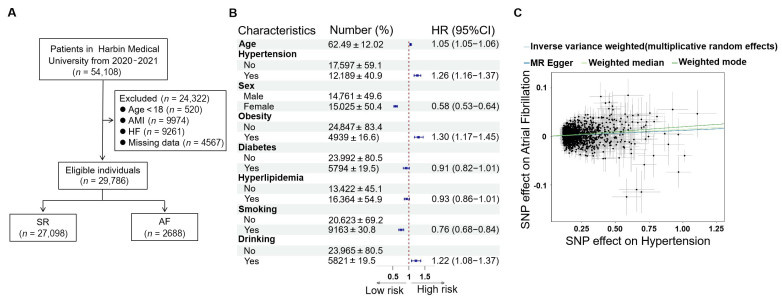
Figure 1. Hypertension as an independent risk factor and causal contributor to atrial fibrillation
(A) Participant flowchart illustrating the design of the observational study based on data from Harbin. A total of 54,108 patients hospitalized at the First Affiliated Hospital of Harbin Medical University between 2020 and 2021 were initially recruited. After excluding patients younger than 18 years, those with acute myocardial infarction (AMI), heart failure (HF), or incomplete clinical data, 29,786 individuals were included in the final analysis. Among these, 27,098 patients were in sinus rhythm, while 2688 were diagnosed with AF. (B) Multivariate logistic regression analysis identifying independent risk factors for AF. (C) Scatter plot depicting the Mendelian randomization analysis assessing the causal relationship between hypertension and AF. SR, sinus rhythm; SNP, single nucleotide polymorphism.

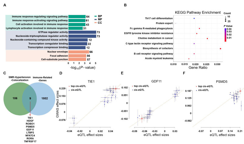
Figure 2. Integration of hypertension eQTL data with immune-related genes
Gene Ontology (GO) enrichment analysis of genes associated with hypertension based on whole blood eQTL data. The analysis identifies significant GO terms across three categories: Biological process (BP), molecular function (MF), and cellular component (CC). (B) KEGG pathway enrichment analysis for genes associated with hypertension based on whole blood eQTL data. (C) Venn diagram showing the overlap between genes identified through Summary-based Mendelian Randomization (SMR) for hypertension colocalization (green) and immune-related genes (blue). (D) Scatter plot showing the correlation between eQTL effect sizes and GWAS effect sizes for the gene TIE1 (ENSG00000006606). (E) Scatter plot showing the correlation between eQTL effect sizes and GWAS effect sizes for the gene GDF11 (ENSG00000135414). (F) Scatter plot showing the correlation between eQTL effect sizes and GWAS effect sizes for the gene PSMD5 (ENSG00000092581).

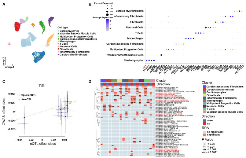
Figure 3. Single-cell sequencing analysis in AF samples
(A) Uniform manifold approximation and projection (UMAP) plot illustrating the clustering of major cell types identified in AF samples, including cardiomyocytes, vascular smooth muscle cells, multipotent progenitor cells, cardiac-associated fibroblasts, macrophages, T cells, neuronal cells, fibroblasts, inflammatory fibroblasts, and cardiac myofibroblasts. (B) Dot plot showing the average expression levels and the proportion of cells expressing selected key genes across the identified cell types. Cardiomyocytes display distinct expression patterns compared with other cell populations. (C) Cis-expression quantitative trait locus (cis-eQTL) analysis of the TIE1 gene (ENSG00000066056) in AF samples. (D) Heatmap depicting enrichment of hallmark gene sets across different cell clusters, with color gradients indicating the direction (up-or down-regulation) and significance of enrichment. Clusters include cardiomyocytes, cardiac-associated fibroblasts, cardiac myofibroblasts, and other cell types. AF, atrial fibrillation.

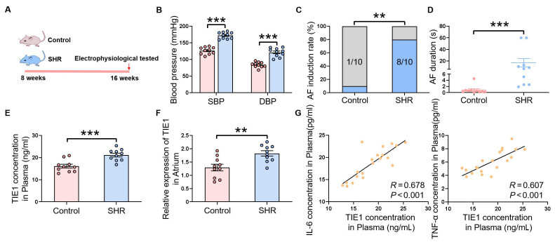
Figure 4. Spontaneously hypertensive rats exhibit increased AF susceptibility and TIE1 expression
(A) Schematic diagram illustrating the experimental design, in which 8-week-old control rats and spontaneously hypertensive rats (SHR) were maintained until 16 weeks of age. (B) Quantification of systolic blood pressure (SBP) and diastolic blood pressure (DBP) in control and SHR groups (N = 10 per group). (C) AF inducibility in control and SHR groups (N = 10 per group). (D) AF duration in control and SHR groups (N = 10 per group). (E) Plasma TIE1 concentrations in control and SHR rats (N = 10 per group). (F) Relative expression levels of TIE1 in atrial tissues from control and SHR rats (N = 10 per group). (G) Correlation between plasma TIE1 levels and IL-6 concentrations in SHR rats. (H) Correlation between plasma TIE1 levels and TNF-α concentrations in SHR rats. Data are presented as mean ± SEM and were analyzed using Student’s t test, Wilcoxon test, or Fisher’s exact test, as appropriate. **P < 0.01; ***P < 0.001. AF, atrial fibrillation.

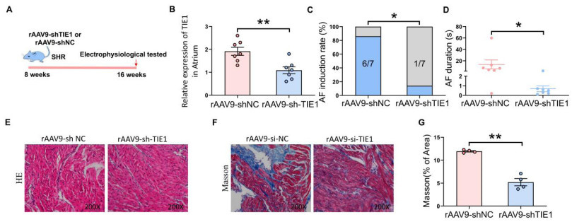
Figure 5. Knockdown of TIE1 reduces atrial fibrillation susceptibility in spontaneously hypertensive rats
(A) Schematic diagram of the experimental design. Spontaneously hypertensive rats were randomly assigned to receive either a negative control vector or an AAV9-mediated TIE1 knockdown vector. AF susceptibility was evaluated 8 weeks after treatment. (B) Relative expression levels of TIE1 in atrial tissues from the rAAV9-shNC and rAAV9-shTIE1 groups (N = 7 per group). (C) AF inducibility in the rAAV9-shNC and rAAV9-shTIE1 groups (N = 7 per group). (D) AF duration in the rAAV9-shNC and rAAV9-shTIE1 groups (N = 7 per group). (E) Representative hematoxylin and eosin (H&E)–stained sections of the left ventricle. Magnification × 200. (F) Representative Masson’s trichrome–stained sections of the left ventricle. Magnification × 200. (G) Quantification of collagen volume fraction in the left ventricle (N = 5 per group). Data are presented as mean ± SEM and were analyzed using Student’s t test, Wilcoxon test, or Fisher’s exact test, as appropriate. *P < 0.05; **P < 0.01. SHR, spontaneously hypertensive rats; AF, atrial fibrillation.
-
[1] Li X, Wu C, Lu J, et al. Cardiovascular risk factors in China: A nationwide population-based cohort study. Lancet Public Health, 2020; 5(12): e672-e681. doi: 10.1016/S2468-2667(20)30191-2 [2] Zhu X, Chen R, Zhang Y, et al. Low ambient temperature increases the risk and burden of atrial fibrillation episodes: A nationwide case-crossover study in 322 Chinese cities. Sci Total Environ, 2023; 880: 163351. doi: 10.1016/j.scitotenv.2023.163351 [3] Chen J, Gao Y, Jiang Y, et al. Low ambient temperature and temperature drop between neighbouring days and acute aortic dissection: A case-crossover study. Eur Heart J, 2022; 43(3): 228-235. doi: 10.1093/eurheartj/ehab803 [4] Zhang Q, Peng L, Hu J, et al. Low temperature and temperature decline increase acute aortic dissection risk and burden: A nationwide case crossover analysis at hourly level among 40,270 patients. Lancet Reg Health West Pac, 2022; 28: 100562. [5] Escolar V, Lozano A, Larburu N, et al. Impact of environmental factors on heart failure decompensations. ESC Heart Fail, 2019; 6(6): 1226-1232. doi: 10.1002/ehf2.12506 [6] Luo Y, Zhang Y, Han X, et al. Akkermansia muciniphila prevents cold-related atrial fibrillation in rats by modulation of TMAO induced cardiac pyroptosis. EBioMedicine, 2022; 82: 104087. doi: 10.1016/j.ebiom.2022.104087 [7] Yang L, Li L, Lewington S, et al. Outdoor temperature, blood pressure, and cardiovascular disease mortality among 23 000 individuals with diagnosed cardiovascular diseases from China. Eur Heart J, 2015; 36(19): 1178-1185. doi: 10.1093/eurheartj/ehv023 [8] Hindricks G, Potpara T, Dagres N, et al. 2020 ESC Guidelines for the diagnosis and management of atrial fibrillation developed in collaboration with the European Association for Cardio-Thoracic Surgery (EACTS): The task force for the diagnosis and management of atrial fibrillation of the European Society of Cardiology (ESC) developed with the special contribution of the European Heart Rhythm Association (EHRA) of the ESC. Eur Heart J, 2021; 42(5): 373-498. doi: 10.1093/eurheartj/ehaa612 [9] Lacoste J L, Szymanski T W, Avalon J C, et al. Atrial fibrillation management: a comprehensive review with a focus on pharmacotherapy, rate, and rhythm control strategies. Am J Cardiovasc Drugs, 2022; 22(5): 475-496. doi: 10.1007/s40256-022-00529-6 [10] Heijman J, Madreiter-Sokolowski C T. Is ageing a modifiable risk factor for atrial fibrillation. Cardiovasc Res, 2024; 120(5): 440-442. doi: 10.1093/cvr/cvae040 [11] Camargo L L, Rios F J, Montezano A C, et al. Reactive oxygen species in hypertension. Nat Rev Cardiol, 2025; 22(1): 20-37. doi: 10.1038/s41569-024-01062-6 [12] Mills K T, Stefanescu A, He J. The global epidemiology of hypertension. Nat Rev Nephrol, 2020; 16(4): 223-237. doi: 10.1038/s41581-019-0244-2 [13] Zhou B, Perel P, Mensah G A, et al. Global epidemiology, health burden and effective interventions for elevated blood pressure and hypertension. Nat Rev Cardiol, 2021; 18(11): 785-802. doi: 10.1038/s41569-021-00559-8 [14] NCD Risk Factor Collaboration (NCD-RisC). Worldwide trends in hypertension prevalence and progress in treatment and control from 1990 to 2019: A pooled analysis of 1201 population-representative studies with 104 million participants. Lancet, 2021; 398(10304): 957-980. doi: 10.1016/S0140-6736(21)01330-1 [15] Deng M, Huang L, Ning B, et al. β-asarone improves learning and memory and reduces acetyl cholinesterase and beta-amyloid 42 levels in APP/PS1 transgenic mice by regulating Beclin-1-dependent autophagy. Brain Res, 2016; 1652: 188-194. doi: 10.1016/j.brainres.2016.10.008 [16] Guo X, Ouyang N, Sun G, et al. Multifaceted intensive blood pressure control model in older and younger individuals with hypertension: A randomized clinical trial. JAMA Cardiol, 2024; 9(9): e241449. [17] Schiffrin E L, Fisher N D L. Diagnosis and management of resistant hypertension. BMJ, 2024; 385: e079108. doi: 10.1136/bmj-2023-079108 [18] Wang C, Liu S, Miao W, et al. Intensive blood pressure control in isolated systolic hypertension: A post hoc analysis of a cluster randomized trial. Lancet Reg Health West Pac, 2024; 48: 101127. [19] Huxley R R, Lopez F L, Folsom A R, et al. Absolute and attributable risks of atrial fibrillation in relation to optimal and borderline risk factors: The Atherosclerosis Risk in Communities (ARIC) study. Circulation, 2011; 123(14): 1501-1508. doi: 10.1161/CIRCULATIONAHA.110.009035 [20] Rahman F, Yin X, Larson M G, et al. Trajectories of risk factors and risk of new-onset atrial fibrillation in the framingham heart study. Hypertension, 2016; 68(3): 597-605. doi: 10.1161/HYPERTENSIONAHA.116.07683 [21] Wu K, Zhu J, Ma Y, et al. Exploring immune related gene signatures and mechanisms linking non alcoholic fatty liver disease to atrial fibrillation through transcriptome data analysis. Sci Rep, 2023; 13(1): 17548. doi: 10.1038/s41598-023-44884-z [22] Chen W, Yang Q, Hu L, et al. Shared diagnostic genes and potential mechanism between PCOS and recurrent implantation failure revealed by integrated transcriptomic analysis and machine learning. Front Immunol, 2023; 14: 1175384. doi: 10.3389/fimmu.2023.1175384 [23] Pozarickij A, Gan W, Lin K, et al. Causal relevance of different blood pressure traits on risk of cardiovascular diseases: GWAS and Mendelian randomisation in 100,000 Chinese adults. Nat Commun, 2024; 15(1): 6265. doi: 10.1038/s41467-024-50297-x [24] Lu J, Wang Z. Mendelian randomisation supports no evidence of the association between asthma and coronary heart disease in East Asians. Eur Respir J, 2024; 64(1): 2400628. doi: 10.1183/13993003.00628-2024 [25] Zhou L, Gao H, Zhang J, et al. Metabolic syndrome and cancer risk: A two-sample Mendelian randomization study of European ancestry. Int J Surg, 2025; 111(1): 311-321. doi: 10.1097/JS9.0000000000001926 [26] Keaton J M, Kamali Z, Xie T, et al. Genome-wide analysis in over 1 million individuals of European ancestry yields improved polygenic risk scores for blood pressure traits. Nat Genet, 2024; 56(5): 778-791. doi: 10.1038/s41588-024-01714-w [27] Roselli C, Chaffin M D, Weng L C, et al. Multi-ethnic genome-wide association study for atrial fibrillation. Nat Genet, 2018; 50(9): 1225-1233. doi: 10.1038/s41588-018-0133-9 [28] Selewa A, Luo K, Wasney M, et al. Single-cell genomics improves the discovery of risk variants and genes of atrial fibrillation. Nat Commun, 2023; 14(1): 4999. doi: 10.1038/s41467-023-40505-5 [29] Hemani G, Zheng J, Elsworth B, et al. The MR-Base platform supports systematic causal inference across the human phenome. Elife, 2018; 7: e34408. doi: 10.7554/eLife.34408 [30] Davies N M, Holmes M V, Davey S G. Reading Mendelian randomisation studies: A guide, glossary, and checklist for clinicians. BMJ, 2018; 362: k601. [31] Zhang J, Liu S, Ding W, et al. Resolution of inflammation, an active process to restore the immune microenvironment balance: A novel drug target for treating arterial hypertension. Ageing Res Rev, 2024; 99: 102352. doi: 10.1016/j.arr.2024.102352 [32] Cheng J, Gu W, Lan T, et al. Single-cell RNA sequencing reveals cell type- and artery type-specific vascular remodelling in male spontaneously hypertensive rats. Cardiovasc Res, 2021; 117(4): 1202-1216. doi: 10.1093/cvr/cvaa164 [33] Nosalski R, Siedlinski M, Denby L, et al. T-Cell-Derived miRNA-214 mediates perivascular fibrosis in hypertension. Circ Res, 2020; 126(8): 988-1003. doi: 10.1161/CIRCRESAHA.119.315428 [34] Ye J, Que B, Huang Y, et al. Interleukin-12p35 knockout promotes macrophage differentiation, aggravates vascular dysfunction, and elevates blood pressure in angiotensin Ⅱ-infused mice. Cardiovasc Res, 2019; 115(6): 1102-1113. doi: 10.1093/cvr/cvy263 [35] Shi H Z, Wang M W, Huang Y S, et al. A telomere-related gene risk model for predicting prognosis and treatment response in acute myeloid leukemia. Heliyon, 2024; 10(11): e31705. doi: 10.1016/j.heliyon.2024.e31705 [36] Li Y, Liu X, Zhao F, et al. Comprehensive analysis of PSMD family members and validation of PSMD9 as a potential therapeutic target in human glioblastoma. CNS Neurosci Ther, 2024; 30(2): e14366. doi: 10.1111/cns.14366 [37] Nijhawan D, Zack T I, Ren Y, et al. Cancer vulnerabilities unveiled by genomic loss. Cell, 2012; 150(4): 842-854. doi: 10.1016/j.cell.2012.07.023 [38] Haertle L, Barrio S, Munawar U, et al. Single-nucleotide variants and epimutations induce proteasome inhibitor resistance in multiple myeloma. Clin Cancer Res, 2023; 29(1): 279-288. doi: 10.1158/1078-0432.CCR-22-1161 [39] Kumar S, Nattamai K J, Hassan A, et al. Repolarization of HSC attenuates HSCs failure in Shwachman-Diamond syndrome. Leukemia, 2021; 35(6): 1751-1762. doi: 10.1038/s41375-020-01054-8 [40] Król W, Machelak W, Zielińska M. GDF11 as a friend or an enemy in the cancer biology. Biochim Biophys Acta Rev Cancer, 2023; 1878(5): 188944. doi: 10.1016/j.bbcan.2023.188944 [41] Kraler S, Balbi C, Vdovenko D, et al. Circulating GDF11 exacerbates myocardial injury in mice and associates with increased infarct size in humans. Cardiovasc Res, 2023; 119(17): 2729-2742. doi: 10.1093/cvr/cvad153 [42] Garrido-Moreno V, Díaz-Vegas A, López-Crisosto C, et al. GDF-11 prevents cardiomyocyte hypertrophy by maintaining the sarcoplasmic reticulum-mitochondria communication. Pharmacol Res, 2019; 146: 104273. doi: 10.1016/j.phrs.2019.104273 [43] Zhao Y, Zhu J, Zhang N, et al. GDF11 enhances therapeutic efficacy of mesenchymal stem cells for myocardial infarction via YME1L-mediated OPA1 processing. Stem Cells Transl Med, 2020; 9(10): 1257-1271. doi: 10.1002/sctm.20-0005 [44] Du G Q, Shao Z B, Wu J, et al. Targeted myocardial delivery of GDF11 gene rejuvenates the aged mouse heart and enhances myocardial regeneration after ischemia-reperfusion injury. Basic Res Cardiol, 2017; 112(1): 7. doi: 10.1007/s00395-016-0593-y [45] Dou F, Wu B, Chen J, et al. PPARα targeting GDF11 inhibits vascular endothelial cell senescence in an atherosclerosis model. Oxid Med Cell Longev, 2021; 2021: 2045259. doi: 10.1155/2021/2045259 [46] Xiang G, Ying K, Jiang P, et al. Growth differentiation factor 11 induces skeletal muscle atrophy via a STAT3-dependent mechanism in pulmonary arterial hypertension. Skelet Muscle, 2022; 12(1): 10. doi: 10.1186/s13395-022-00292-x [47] Du J, Liu P, Zhou Y, et al. The mechanosensory channel PIEZO1 functions upstream of angiopoietin/TIE/FOXO1 signaling in lymphatic development. J Clin Invest, 2024; 134(10): e176577. doi: 10.1172/JCI176577 [48] Brouillard P, Murtomäki A, Leppänen V M, et al. Loss-of-function mutations of the TIE1 receptor tyrosine kinase cause late-onset primary lymphedema. J Clin Invest, 2024; 134(14): e173586. doi: 10.1172/JCI173586 [49] Akwii R G, Mikelis C M. Targeting the Angiopoietin/Tie pathway: Prospects for treatment of retinal and respiratory disorders. Drugs, 2021; 81(15): 1731-1749. doi: 10.1007/s40265-021-01605-y [50] Griffin M E, Sorum A W, Miller G M, et al. Sulfated glycans engage the Ang-Tie pathway to regulate vascular development. Nat Chem Biol, 2021; 17(2): 178-186. doi: 10.1038/s41589-020-00657-7 -
 fzm-6-2-86_ESM.docx

-

 下载:
下载:
 点击查看大图
计量
- 文章访问数: 124
- HTML全文浏览量: 65
- PDF下载量: 3
- 被引次数: 0